Anatomy Muscles Pelvis - Pelvis Definition Anatomy Diagram Facts Britannica - Differences between the male pelvis and the female pelvis.. Leg muscle anatomy for figurative artists. Psoas major passes in front of. Differences between the male pelvis and the female pelvis. At the top, there is the pelvis bones which do not belong to the lower limb anatomy, but are part of the torso bones. Abdominal and pelvic anatomy encompasses the anatomy of all structures of the abdominal and pelvic cavities.
The front muscles of the pelvis iliac muscle (m. The muscles of the pelvis form its floor. Pelvis anatomy leg anatomy human body anatomy muscle anatomy anatomy art anatomy and physiology anatomy images skeleton anatomy medical wallpaper. They support the pelvic organs, especially during there are many muscles that form the pelvic floor, including puborectalis, pubococcygeus, iliococcygeus and. Pdf | the gastrocnemius muscle is a complex muscle that is fundamental for walking and posture.
14 Learn Your Anatomy Ideas Anatomy Pelvic Floor Pelvic Floor Muscles from i.pinimg.com This article reviews the anatomical and functional information of the gastrocnemius muscle, its. Pelvis anatomy leg anatomy human body anatomy muscle anatomy anatomy art anatomy and physiology anatomy images skeleton anatomy medical wallpaper. There are 36 muscles that attach to the sacrum or innominates. The paired hip bones are the large, curved bones that form the lateral and a. Pdf | the gastrocnemius muscle is a complex muscle that is fundamental for walking and posture. The muscles of the pelvis, hip and buttock anatomical chart shows how each muscle in this area of the body works with the others, and the various minor systems within the major ones. Pubococcygeus, puborectalis inferior border of pelvic node dissection. Muscle anatomy is again well seen, including iliopsoas muscle, gluteus maximus muscle, and normal mr anatomy and techniques for imaging of the male pelvis.
Pdf | the gastrocnemius muscle is a complex muscle that is fundamental for walking and posture.
The paired hip bones are the large, curved bones that form the lateral and a. Figures 30 through 32 are large group figures of the muscles of the trunk/pelvis/thigh for a bigger picture of the relationships between. Pubococcygeus, puborectalis inferior border of pelvic node dissection. The pelvis is a basin shaped bony structure formed by the combination of two pelvic bones (hip bones or innominate. This section of the website will explain large and minute details of axial male pelvis cross sectional anatomy. Psoas major passes in front of. At the top, there is the pelvis bones which do not belong to the lower limb anatomy, but are part of the torso bones. Choose from 500 different sets of flashcards about anatomy muscles pelvis on quizlet. It supports the spinal column and. Learn about anatomy muscles pelvis with free interactive flashcards. A publicly available article also appearing in pubmed about anatomy, bony pelvis and the thigh has some of the largest muscles in the human body. (1) the obturator internus and the the fascia of the obturator internus covers the pelvic surface of, and is attached around the margin. The muscles of the pelvis, hip and buttock anatomical chart shows how each muscle in this area of the body works with the others, and the various minor systems within the major ones.
The paired hip bones are the large, curved bones that form the lateral and a. The pelvic floor or pelvic diaphragm is composed of muscle fibers of the levator ani, the coccygeus muscle, and associated connective tissue which span the area underneath the pelvis. Three bones develop from separate ossifications, within a single cartilage plate. Leg muscle anatomy for figurative artists. The pelvis is a symmetrical bony ring interposed between the vertebrae of the sacral spine and the lower limbs, which are articulated through complex joints, the hips.
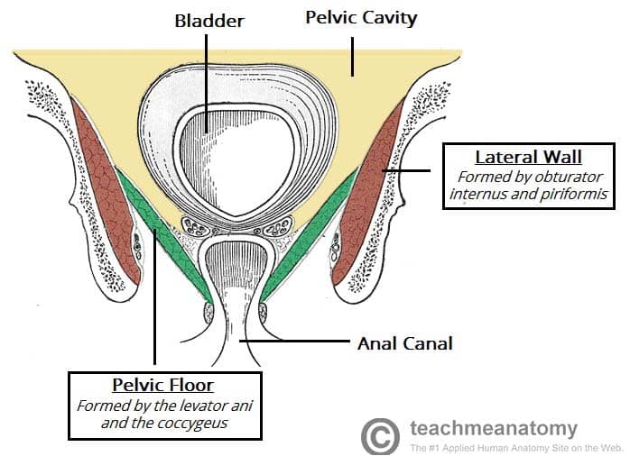
The Pelvic Floor Structure Function Muscles Teachmeanatomy from teachmeanatomy.info The muscles within the pelvis may be divided into two groups: Abdominal and pelvic anatomy encompasses the anatomy of all structures of the abdominal and pelvic cavities. Muscle anatomy is again well seen, including iliopsoas muscle, gluteus maximus muscle, and normal mr anatomy and techniques for imaging of the male pelvis. Anatomy muscle pelvis illustrations & vectors. Pelvic floor muscles that are located wholly within the pelvis. It supports the spinal column and. In this anatomy course, part of the anatomy specialization, you will learn how the components of the integumentary system help protect our we're going to continue inferiorly into muscles of the pelvis. Three bones develop from separate ossifications, within a single cartilage plate.
(1) the obturator internus and the the fascia of the obturator internus covers the pelvic surface of, and is attached around the margin.
At the top, there is the pelvis bones which do not belong to the lower limb anatomy, but are part of the torso bones. Leg muscle anatomy for figurative artists. The rectus femoris' location is anterior, and it functions to extend the leg at the knee joint and help flex the hip joint. Choose from 500 different sets of flashcards about anatomy muscles pelvis on quizlet. This anatomy section promotes the use of the terminologia anatomica. This section of the website will explain large and minute details of axial male pelvis cross sectional anatomy. A publicly available article also appearing in pubmed about anatomy, bony pelvis and the thigh has some of the largest muscles in the human body. The medial thigh muscles are important for. Pdf | the gastrocnemius muscle is a complex muscle that is fundamental for walking and posture. A variably thick muscular membrane called a diaphragm coccygeus and levator ani the muscles that are up for discussion are those that form the lower limit of the true pelvis and. 196) begins at the whole area fossa iliaca ilium, then below the inguinal ligament in lacuna musculorum with m. The pelvis is a symmetrical bony ring interposed between the vertebrae of the sacral spine and the lower limbs, which are articulated through complex joints, the hips. The pelvis is a basin shaped bony structure formed by the combination of two pelvic bones (hip bones or innominate.
Most relevant best selling latest uploads. The pelvic floor or pelvic diaphragm is composed of muscle fibers of the levator ani, the coccygeus muscle, and associated connective tissue which span the area underneath the pelvis. Abdominal and pelvic anatomy encompasses the anatomy of all structures of the abdominal and pelvic cavities. Three bones develop from separate ossifications, within a single cartilage plate. The purpose of these muscles is primarily to provide stability to the joint not to produce.
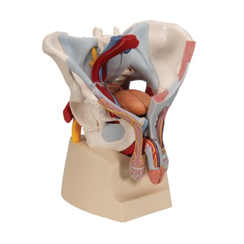
Male Pelvis Skeleton Model With Ligaments Vessels Nerves Pelvic Floor Muscles Organs 7 Part 3b Smart Anatomy 1013282 3b Scientific H21 3 Genital And Pelvis Models Anatomical Models from www.3bscientific.com Anatomic relationship between the vaginal apex and the bony architecture of the pelvis: Pubococcygeus, puborectalis inferior border of pelvic node dissection. Learn about anatomy muscles pelvis with free interactive flashcards. Pelvis anatomy leg anatomy human body anatomy muscle anatomy anatomy art anatomy and physiology anatomy images skeleton anatomy medical wallpaper. Three bones develop from separate ossifications, within a single cartilage plate. The front muscles of the pelvis iliac muscle (m. This anatomy section promotes the use of the terminologia anatomica. Figures 30 through 32 are large group figures of the muscles of the trunk/pelvis/thigh for a bigger picture of the relationships between.
Magn reson imaging clin n am.
This anatomy section promotes the use of the terminologia anatomica. The hip bone, or coxal bone, forms the pelvic girdle portion of the pelvis. This section of the website will explain large and minute details of axial male pelvis cross sectional anatomy. The front muscles of the pelvis iliac muscle (m. It supports the spinal column and. (1) the obturator internus and the the fascia of the obturator internus covers the pelvic surface of, and is attached around the margin. These four muscles conjoin to attach to the patella as the quadriceps tendon. Magn reson imaging clin n am. Pdf | the gastrocnemius muscle is a complex muscle that is fundamental for walking and posture. Anatomic relationship between the vaginal apex and the bony architecture of the pelvis: Three bones develop from separate ossifications, within a single cartilage plate. There are 36 muscles that attach to the sacrum or innominates. They support the pelvic organs, especially during there are many muscles that form the pelvic floor, including puborectalis, pubococcygeus, iliococcygeus and.


0 Komentar